A growth scan is not a weighing machine and a Doppler is not a second opinion on it — here is what each one measures, why the gap between two scans matters as much as the scans, and when the two together change what happens next.
By the third trimester, most of what a pregnancy scan can settle has been settled. The dates were fixed early, the 11-to-14-week scan has done its screening, the anomaly scan has checked the baby’s structure. One question can only be answered late: is this baby growing as it should, and can the placenta still support it?
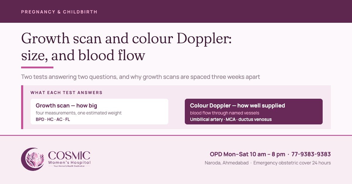
A growth scan and a colour Doppler answer it. They are ordered together often enough that most people assume they are one test. They are not: one measures size, the other measures blood flow, and it is the second that usually decides what happens next.
What a growth scan measures
Four measurements are taken: the biparietal diameter across the head, the head circumference, the abdominal circumference at the level of the stomach and liver, and the femur length. A formula combines them into an estimated fetal weight, which is plotted as a centile against the dates.
The abdominal circumference matters more than its share of the formula suggests. When a placenta begins to underperform, the abdomen is usually where it shows first — and the published definitions of a small baby use it on its own as readily as the estimated weight.
Why the report says “estimated”
A systematic review of the eleven formulae in use found significant error in all of them, with ultrasound most often overestimating the weight. The error comes from two places: measuring a large baby late in pregnancy is genuinely difficult, and the rest is operator dependent — experience, training and audit.
So a figure in grams is the midpoint of a range. Told that her baby is 2,400 g at 34 weeks, the honest reading is “around 2,400 g, and possibly a little less than that”. Nothing is decided on one number.
Three weeks between growth scans, not one
Growth is a velocity — it can only be seen by comparing two measurements. Take them too close together and the margin of error in each swamps the real growth between them, and the comparison starts producing growth restriction that is not there.
The RCOG guideline puts a figure on it: two measurements used to judge growth velocity “should be at least 3 weeks apart to minimise false-positive rates for diagnosing FGR”. This is the single most useful thing to know before asking for an early repeat scan.
Small, and small for a reason
Two babies can weigh the same at 34 weeks and be in entirely different situations. One is simply small — small parents, small baby, growing steadily along its own line. The other has stopped getting what it needs and is falling away from the line it was on. Size alone cannot separate them, which is why the Doppler exists.
The consensus definition does. Small for gestational age is size below a threshold, usually under the 10th centile. Fetal growth restriction means the baby has not reached its own potential, and its criteria use blood flow alongside size, split at 32 weeks.
| Before 32 weeks | From 32 weeks | |
|---|---|---|
| Size alone is enough if | AC or estimated weight below the 3rd centile, or no end-diastolic flow in the umbilical artery | AC or estimated weight below the 3rd centile |
| Otherwise, two of these | AC or weight under the 10th centile · uterine artery PI over the 95th · umbilical artery PI over the 95th | AC or weight under the 10th centile · growth crossing more than two quartiles · cerebroplacental ratio under the 5th, or umbilical artery PI over the 95th |
Delphi consensus criteria, endorsed by ISUOG 2020. PI is the pulsatility index, a measure of resistance to flow.
What a colour Doppler is actually looking at
A Doppler is not a clearer picture. It is a waveform — the shape of blood flow through one named vessel across a heartbeat — and each vessel answers a different question.
| Vessel | What the waveform shows | When it is used |
|---|---|---|
| Uterine artery | How well the mother’s circulation has adapted to the placenta | At 20 to 24 weeks in a woman already at risk. An abnormal result means serial growth scans and umbilical artery Doppler from 26 to 28 weeks |
| Umbilical artery | Resistance inside the placenta itself | The main surveillance tool once a baby measures small |
| Middle cerebral artery, and the cerebroplacental ratio | Whether blood is being redirected to protect the brain | At term with a normal umbilical artery, to help time delivery. Before term it is not used for that |
| Ductus venosus | How the baby’s own heart is coping | In growth restriction before 32 weeks, with computerised CTG, to time delivery |
The umbilical artery does most of the work, and its evidence is unusually clean. Across 16 trials and 10,225 high-risk pregnancies, umbilical artery Doppler brought perinatal deaths down from 1.7% to 1.2%, with fewer inductions and fewer caesareans — pooled international trial data, not this centre’s own figures. Few obstetric tests have a mortality benefit behind them.
The changes come in order, and each one has a response
Placental failure is not a switch. It shows as a sequence, and knowing the sequence is what turns a worrying scan into a plan with dates on it.
After 32 weeks the usual pattern is growth measured fortnightly and Doppler weekly. An abnormal cerebroplacental ratio is confirmed on a repeat within 24 hours before anything is decided on it, because a single reading has a real false-positive rate.
How it is decided who needs one
These are indication-led tests, and the guidelines are unambiguous about why. Routine measurement of abdominal circumference or estimated weight in the third trimester, in women with no risk factor, “does not reduce the incidence of a SGA neonate nor does it improve perinatal outcome”. Routine Doppler in an unselected population gets the same verdict across five trials and 14,185 women, and the WHO antenatal recommendations say as much.
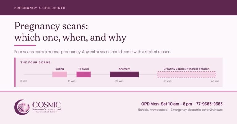
At Cosmic, one growth scan with colour Doppler is done routinely at 32 to 34 weeks. Any further growth scans and Dopplers after that are decided on the findings and the history.
What changes outcomes is doing these tests for a reason — a previous small baby or stillbirth, raised blood pressure or pre-eclampsia, diabetes, twins, reduced movements, a fundal height not keeping pace, an abnormal uterine artery Doppler, or a placenta that needed a third-trimester look anyway. That is high-risk pregnancy care, in which a growth scan is one instrument rather than a date on the calendar. Prevention sits earlier still: where there is real risk of pre-eclampsia and a small baby, low-dose aspirin helps, and the benefit is seen when it is started at or before 16 weeks.
If the scan or the Doppler shows something
The first step is a targeted repeat, not a referral — measurements retaken, the Doppler repeated in the vessels that matter, the picture read against the dates and the earlier scans. Most of the time it ends there.
Where the finding stands up, confirmed growth restriction or a Doppler finding that puts the timing of delivery in play is referred to an expert fetal medicine specialist for the detailed assessment and counselling — while the pregnancy continues to be looked after here, with the delivery planned around what that assessment says.
At Cosmic
- Growth scans and colour Doppler are performed and reported by Dr. Rahul Khatri and Dr. Bhargavi Khatri themselves — scan and clinical decision sit with the same person.
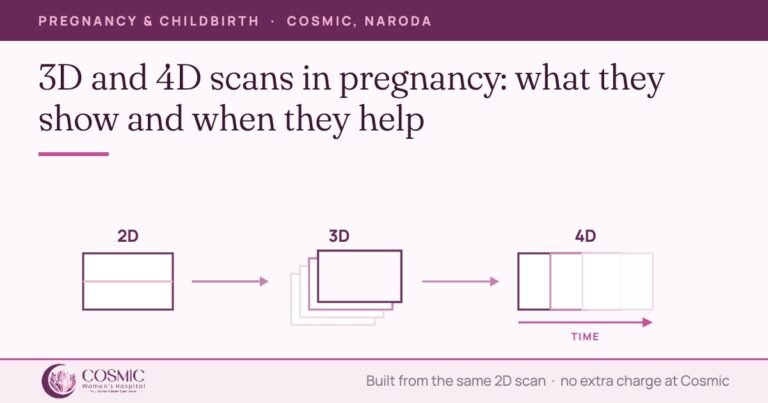
- Obstetric ultrasound is Rs 1,200 and a colour Doppler is Rs 1,500, covering the vessels the findings call for — umbilical artery, middle cerebral artery and ductus venosus. 3D and 4D imaging is available within an indicated scan at no additional charge.
- A growth scan with colour Doppler is done routinely at 32 to 34 weeks, with the fetal weight estimated by the Hadlock formula. Any further growth scans and Dopplers after that are decided on the findings and the history.
- Antenatal visits run 2-weekly through the eighth month and weekly through the ninth, which is when a growth assessment is usually fitted.
- OPD Monday to Saturday, 10 am to 8 pm, with obstetric emergency cover 24 hours.
What the law requires at every pregnancy scan
Prenatal sex determination is not done at Cosmic Women’s Hospital and is prohibited by law under the Pre-conception and Pre-natal Diagnostic Techniques (Prohibition of Sex Selection) Act, 1994.
The sex of the baby is not disclosed at any scan, by words, signs or in any other manner whatsoever. A Form F is completed and signed for every obstetric scan, the written indication is recorded, and records are maintained as the Act requires. The notice to this effect is displayed at the ultrasound room in English and in Gujarati. These obligations apply to a growth scan and a colour Doppler exactly as to any other prenatal ultrasound.
The scans of pregnancy work as a sequence, not as separate appointments. Our guide to pregnancy scans covers the whole sequence, and the obstetric service it sits inside is described on our maternity hospital page.
Sources
- Lees CC, Stampalija T, Baschat AA, et al. ISUOG Practice Guidelines: diagnosis and management of small-for-gestational-age fetus and fetal growth restriction. Ultrasound Obstet Gynecol 2020;56:298–312.
- ISUOG Practice Guidelines (updated): use of Doppler velocimetry in obstetrics. Ultrasound Obstet Gynecol 2021;58:331–339.
- Royal College of Obstetricians and Gynaecologists. The Investigation and Management of the Small-for-Gestational-Age Fetus. Green-top Guideline No. 31, 2nd edition, 2013.
- Milner J, Arezina J. The accuracy of ultrasound estimation of fetal weight in comparison to birth weight: a systematic review. Ultrasound 2018;26(1):32–41.
- World Health Organization. WHO recommendations on antenatal care, 2016 — recommendations B.2.4 and B.2.5.
- National Institute for Health and Care Excellence. Antenatal care. NICE guideline NG201, 2021 — Schedule of antenatal appointments.
- Pre-conception and Pre-natal Diagnostic Techniques (Prohibition of Sex Selection) Act, 1994 and Rules.
Written by the clinical team at Cosmic Women’s Hospital & IVF Center, Naroda, Ahmedabad.
Medically reviewed by Dr. Rahul Khatri
MBBS, MS & DNB Obgy, FMAS
Consulting Obgyn, Laparoscopic & Robotic Surgeon, Director
Last reviewed: 2 October 2026
General information about obstetric ultrasound, not a substitute for an individual consultation. Every decision here is taken for each pregnancy on its own findings.
Booking a growth scan or a Doppler
3rd Floor, The Capital Corporate, Nr. Naroda Muktidham, Naroda, Ahmedabad 382330. OPD Monday to Saturday, 10 am to 8 pm. Call 77-9383-9383.
3rd Floor, The Capital Corporate, Nr. Naroda Muktidham, Naroda, Ahmedabad 382330. OPD Monday to Saturday, 10 am to 8 pm. Call 77-9383-9383.
Book a consultation
Tell us how we can help and we will call you back.

Cosmic Women's Hospital
Cosmic Women’s Hospital & IVF Center is a registered Level 2 ART clinic in Naroda, Ahmedabad — obstetrics, gynaecological surgery, IVF and ICSI, with ultrasound in-house and 24-hour emergency obstetric cover. Articles here are written by the clinical team and medically reviewed by Dr. Rahul Khatri or Dr. Bhargavi Khatri before publication.