The 11-to-14-week scan has a deadline, measures one thing to a tenth of a millimetre, and gives you a risk rather than an answer — here is what it settles, what it cannot, and what Indian law says about it.
Why this scan has a deadline
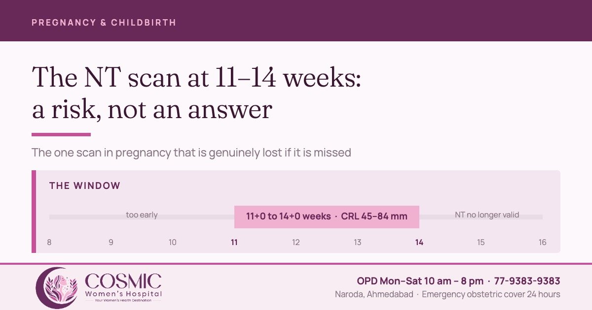
Most scans in pregnancy can be done a few days early or a few days late without much being lost. The nuchal translucency scan cannot. It is performed between 11+0 and 14+0 weeks, when the crown–rump length measures 45 to 84 mm. Before 11 weeks the thin layer of fluid at the back of the fetal neck is too small to measure dependably; after 14 weeks it settles and the measurement stops carrying the meaning the calculation depends on. The NHS schedule in the UK, for the same reason, books the appointment between 11+2 and 14+1 weeks.
This is the one scan appointment in pregnancy that is genuinely lost if it is missed, so the date of the first visit matters more than most women expect — particularly if the last period is uncertain or there have been earlier pregnancy losses.
Sex determination is not done, and cannot be
Prenatal sex determination is not done at this hospital and is prohibited by law under the Pre-conception and Pre-natal Diagnostic Techniques (Prohibition of Sex Selection) Act, 1994.
The Act places specific obligations on every registered ultrasound facility. A Form F is completed for every prenatal diagnostic procedure and signed by the person conducting it; the medical reason for the scan is recorded in writing; written consent is taken; records are kept for two years; and a notice stating that sex determination is not done here is displayed prominently. The Act forbids disclosure of the sex of the fetus “by words, signs or in any other manner whatsoever”.
The penalties fall on both sides — up to three years’ imprisonment for the practitioner on a first offence, and up to three years and a fine of up to Rs 50,000 for the person who seeks it. Please do not ask the sonologist, and do not ask the staff.
What is actually measured
The nuchal translucency is a normal, fluid-filled space at the back of the fetal neck. Every fetus has one. What matters is how thick it is for the size of the fetus — and that is a measurement made under strict conditions, not an impression formed on screen.
The image is taken in a mid-sagittal section, magnified so that only the head and upper chest are on screen. The neck must be in a neutral position, because flexing or extending it changes the number, and the fetus must be clearly separate from the amnion so that the right space is being measured. Cross calipers are placed on the echogenic margins at the widest point, on a machine measuring in 0.1 mm steps. Three measurements are taken on three separate images, and the largest is the one used in the calculation.
That sequence is why the scan takes the time it does, and why a hurried NT measurement is worth less than none at all. The same image also allows the nasal bone to be assessed, and flow across the tricuspid valve and in the ductus venosus can be added as further markers — which ISUOG notes improve the screening and also “require additional skills for reliable assessment”.
A risk, not a diagnosis
The NT measurement is combined with maternal age, the gestational age from the same scan, and two blood markers taken at the same time — PAPP-A and free beta-hCG. Software turns that into a number: a probability, such as 1 in 1,200 or 1 in 60, that this pregnancy is affected by Down syndrome (trisomy 21) or by trisomy 18 or 13. It does not say yes and it does not say no.
How good it is depends entirely on what is combined with what.
| Route | What it involves | Trisomy 21 found | Called screen-positive |
|---|---|---|---|
| NT measurement alone | Scan only, adjusting the age-related risk | about 70% | — |
| First-trimester combined | NT + PAPP-A + free beta-hCG at 11–14 weeks | 82–87% | 5% |
| Quadruple marker | Blood only, second trimester | 81% | 5% |
| Cell-free DNA | Maternal blood sample, from 10 weeks | 99% | 2–4% |
Those rates are as published in ACOG Practice Bulletin 226; ISUOG reports combined first-trimester screening detecting 90% of trisomy 21, 97% of trisomy 18 and 92% of trisomy 13 at 96% specificity. The figure worth holding on to is the first row: the scan on its own finds roughly seven cases in ten. The blood markers are not an optional extra, and they are drawn at the same visit because they only work inside the same window.
If the result comes back high risk
A high-risk result is not a diagnosis, and at younger maternal ages it is quite often not even a correct prediction. Of women whose cell-free DNA test reads positive for trisomy 21 at 10 weeks, ACOG puts the proportion who really are carrying an affected pregnancy at 38–80% at age 20, and at 91–99% at age 40 — the same laboratory result, the same words on the report, and a very different meaning.
Cell-free DNA also fails to give a result in about 3% of samples, and ACOG notes that this group carries a higher chance of aneuploidy than the population it came from — a no-call is a reason to be seen again, not a reason to relax. And for all its accuracy, ACOG is explicit that “cell-free DNA testing is not equivalent to diagnostic testing”. Only a sample of the pregnancy itself tests the chromosomes.
| Test | When | What it gives | Procedure-related miscarriage |
|---|---|---|---|
| Cell-free DNA | From 10 weeks | A refined screening result. Needs confirmation before any decision | None — it is a blood test |
| Chorionic villus sampling | From about 11 weeks | A diagnosis from placental tissue | 0.22% |
| Amniocentesis | From 15 weeks | A diagnosis from amniotic fluid | 0.11% |
Those two figures come from a meta-analysis of 42,716 amniocenteses and 8,899 CVS procedures, and they are far lower than the one-in-a-hundred most patients have been told. In the same analysis, the background miscarriage rate in comparable women who had no procedure was 0.67% and 1.79% — so most losses after an amniocentesis would have happened anyway. The authors conclude the risks “are much lower than are currently quoted”. That does not make an invasive test trivial, but the decision should rest on what the couple wants to know, not on a number remembered from a decade ago.
Four things this scan settles besides the number
Patients think of this appointment as the Down syndrome test. Clinically it is doing four other jobs, any one of which would justify it.
- Dates. The crown–rump length at this stage is the most precise dating measurement in pregnancy — accurate to within five days either way in 95% of cases. Growth percentiles, the timing of delivery and the whole question of whether a pregnancy is post-dates all rest on the date fixed here. Above 84 mm, dating moves to head circumference and loses precision.
- How many, and how they share. In a twin pregnancy this is the window in which chorionicity and amnionicity can still be determined reliably, and that one finding changes the entire plan.
- Early anatomy. A structured survey checks the skull outline and calcification, the two brain halves with the choroid plexuses filling the ventricles, a four-chamber heart beating regularly inside the chest, the stomach, bladder, an intact abdominal wall, and four limbs with three segments each.
- A first look at the heart. ISUOG notes the NT is increased in up to 40% of fetuses with a major cardiac abnormality — which is why a raised measurement leads to a detailed look at the heart later even when the chromosomes are normal.
What a normal result does not rule out
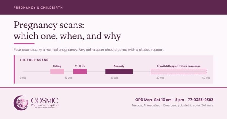
ISUOG is careful about this and so should we be: a significant proportion of structural anomalies can be found at 11 to 14 weeks, but “many major malformations may develop later in pregnancy or may not be detected even with appropriate equipment and in the most experienced of hands”. A reassuring NT scan is not a clean bill of health. The mid-trimester anomaly scan between 18 and 24 weeks remains part of the plan, and the WHO recommends at least one scan before 24 weeks for every pregnancy. Which scans a pregnancy needs, and when, is set out in our guide to pregnancy scans.
At Cosmic
Obstetric ultrasound is done in our own scan room at Naroda, so the 11-to-14-week appointment, the blood samples taken with it and the consultation that follows all happen in one visit. The hospital is a registered facility under the PC&PNDT Act, Form F is completed for every scan, and a female attendant is present throughout. Antenatal visits then run three-weekly in the first trimester, four-weekly in the second, and closer from the seventh month — the schedule is set out in full on high-risk pregnancy care and maternity care at Naroda.
| Item | Charge |
|---|---|
| Consultation, new obstetric or gynaecological case | Rs 500 |
| Follow-up consultation | Rs 300 |
| Obstetric ultrasound | Rs 1,200 |
| Colour Doppler | Rs 1,500 |
The combined test is done in full here, in one visit: the scan, the blood markers and the consultation. The blood markers are quoted before anything is done. If a cell-free DNA (NIPT) test or an invasive test (CVS or amniocentesis) is needed, you are referred to an expert fetal medicine specialist, and your pregnancy continues to be looked after here. OPD Monday to Saturday, 10 am to 8 pm, with 24-hour emergency obstetric cover. To book the scan inside its window, call 77-9383-9383.
Four questions worth asking at the appointment
- How many completed weeks and days am I today — from my dates, or from this scan?
- Are the blood markers being drawn today, or is the NT being reported on its own?
- What is the risk figure, and against what cut-off is it being called low or high?
- If it comes back high risk, what is offered next, and when does that window close?
Sources
- ISUOG Practice Guidelines (updated): performance of 11–14-week ultrasound scan. Ultrasound in Obstetrics & Gynecology 2023;61:127–143.
- ISUOG Practice Guidelines: performance of first-trimester fetal ultrasound scan. Ultrasound in Obstetrics & Gynecology 2013;41:102–113.
- ACOG Practice Bulletin No. 226: Screening for Fetal Chromosomal Abnormalities. Obstetrics & Gynecology 2020;136:e48–e69.
- Akolekar R, Beta J, Picciarelli G, Ogilvie C, D’Antonio F. Procedure-related risk of miscarriage following amniocentesis and chorionic villus sampling: a systematic review and meta-analysis. Ultrasound in Obstetrics & Gynecology 2015;45:16–26.
- NICE NG201, Antenatal care (2021) — Schedule of antenatal appointments.
- WHO recommendations on antenatal care for a positive pregnancy experience (2016), recommendation B.2.4.
- The Pre-conception and Pre-natal Diagnostic Techniques (Prohibition of Sex Selection) Act, 1994 and Rules — Ministry of Health and Family Welfare handbook.
Written by the clinical team at Cosmic Women’s Hospital & IVF Center, Naroda, Ahmedabad.
Medically reviewed by Dr. Rahul Khatri — MBBS, MS & DNB Obgy, FMAS. Consulting Obgyn, Laparoscopic & Robotic Surgeon, Director.
Reviewed 28 September 2026. This article is general information about a screening test and is not a substitute for a consultation. Prenatal sex determination is not done at this hospital and is prohibited by law under the PC&PNDT Act, 1994.
Book a consultation
Tell us how we can help and we will call you back.
Cosmic Women's Hospital
Cosmic Women’s Hospital & IVF Center is a registered Level 2 ART clinic in Naroda, Ahmedabad — obstetrics, gynaecological surgery, IVF and ICSI, with ultrasound in-house and 24-hour emergency obstetric cover. Articles here are written by the clinical team and medically reviewed by Dr. Rahul Khatri or Dr. Bhargavi Khatri before publication.