The four scans that actually decide something, the weeks each one belongs in, what each can and cannot find — and why a pregnancy going normally needs fewer scans than most people are given.
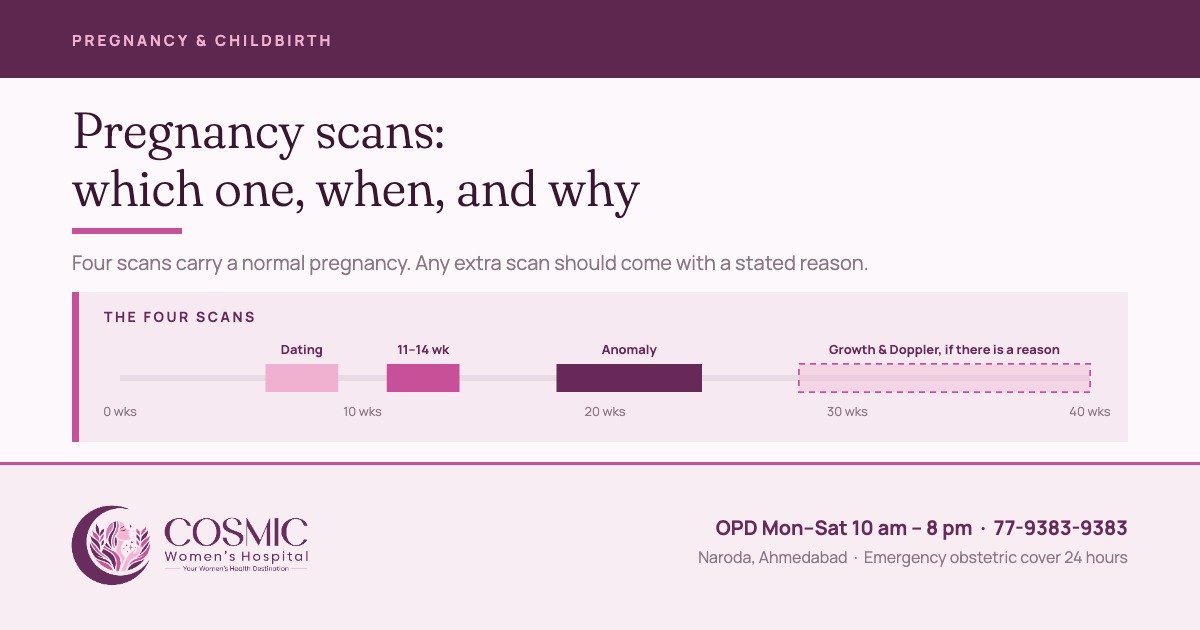
How many scans does a pregnancy need? Ask three hospitals in Ahmedabad and you will get three answers, and the range is wide enough that it is worth understanding rather than simply accepting. Some women are scanned once a month. Some are scanned four times in nine months and told that is enough. Both cannot be right.
If this is your first visit to us, what a first consultation involves is set out separately, and the full range of services is on the Naroda gynaecology page.
This page sets out the four scans that actually decide something, when each one is done, what each can and cannot tell you, and why a normal pregnancy needs fewer of them than most people expect.
The four scans that decide something
| Scan | When | What it settles |
|---|---|---|
| Dating and viability | About 6–9 weeks, where it is needed | That the pregnancy is in the uterus, that there is a heartbeat, how many, and roughly how far along |
| The 11–14 week scan | 11+0 to 14+0 weeks | The dating that the whole pregnancy will run on, nuchal translucency, the number of babies and their chorionicity, and a first look at the anatomy |
| The anomaly scan | 18–24 weeks | The detailed structural survey, and where the placenta lies |
| Growth and Doppler | 32–34 weeks routinely at Cosmic, and again when there is a reason | Whether the baby is growing as expected and how the blood flow looks |
Why the first scan sets the date for everything after it
Gestational age is fixed early and then left alone, because early measurement is far more precise than late measurement. The crown–rump length — the length of the baby from head to bottom — dates a pregnancy to within five days either way in 95 per cent of cases.1 Once that measurement passes 84 mm, the head circumference becomes the more reliable one.1
This matters more than it sounds. The date set at the first scan is the date used to decide whether the baby is small at 32 weeks, whether you are overdue at 41, and whether an induction is offered, and it feeds into how the route of delivery is decided. A pregnancy dated by a third-trimester scan is dated badly, and the errors are measured in weeks rather than days.
The 11 to 14 week scan
Done between 11+0 and 14+0 weeks, with the crown–rump length between 45 and 84 mm.2 It carries more weight than any other scan in the pregnancy, because it does four jobs at once: it fixes the dates, it measures the nuchal translucency for the aneuploidy risk assessment, it establishes in a twin pregnancy whether the babies share a placenta, and it gives an early look at the anatomy. A good proportion of severe structural anomalies can already be seen at this stage.2 What it measures, how accurate it is and what a high-risk result means are set out in our NT scan guide.
The chorionicity point is the one families most often miss. In twins, whether the babies share a placenta changes the entire monitoring schedule for the rest of the pregnancy, and it can be established easily in the first trimester and only with difficulty later.
The anomaly scan
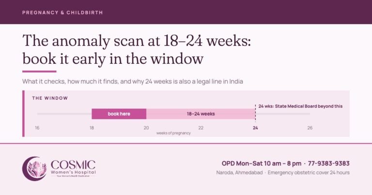
Performed between 18 and 24 weeks.3 This is the long one, and the one worth arriving unhurried for. What is checked, and why booking it for 18 to 20 weeks matters in India, is in our anomaly scan guide.
It is a careful examination, not a guarantee, and it is more honest to say so before the scan than afterwards. The UK screening programme, which audits its own performance, reports that the scan finds around 9 in 10 babies with open spina bifida and around half of those with heart conditions.4 These are overall published figures for a national programme, not this centre's own detection rates. Some conditions are not visible on any scan. Some appear only later in pregnancy. A normal anomaly scan is genuine reassurance, and it is not a certificate.
The scan also records where the placenta is lying, which is what decides whether a repeat scan is needed later to see if it has moved away from the cervix.
Growth scans and Doppler in the last three months
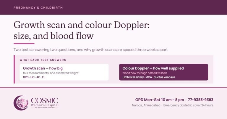
Here is where the practice between hospitals diverges most, and where the evidence is clearest. What each test measures, and why growth scans are spaced at least three weeks apart, is set out in our growth scan and Doppler guide.
In a pregnancy that is going normally, routine scanning in the third trimester does not improve the outcome. The RCOG puts it as a Grade A recommendation: routine measurement of abdominal circumference or estimated fetal weight in the third trimester neither reduces the number of small babies born nor improves perinatal outcome, on the evidence of eight trials and 27,024 women.5 The same guideline finds no conclusive benefit from routine umbilical artery Doppler in a low-risk population, across five trials and 14,185 women.5 The World Health Organization reaches the same conclusion and does not recommend routine Doppler in pregnancy.6
At Cosmic, one growth scan with colour Doppler is done routinely at 32 to 34 weeks. Any further growth scans and Dopplers after that are decided on the findings and the history.
In a pregnancy that is being watched — a small baby, raised blood pressure, a previous growth-restricted baby, a medical condition — see high-risk pregnancy care — umbilical artery Doppler is a different instrument altogether. Across 16 trials and 10,225 babies it reduced perinatal deaths from 1.7 per cent to 1.2 per cent, and reduced inductions and caesareans with it.5 That is why it is the primary surveillance tool once a baby is suspected of being small.
What this means for you. If we are not scanning you every month, that is a clinical decision, not an economy. If we start scanning you every two or three weeks, that is also a clinical decision and you should ask what changed. Either way the answer should be a sentence you can repeat to your family, not “just to check”.
3D and 4D scans
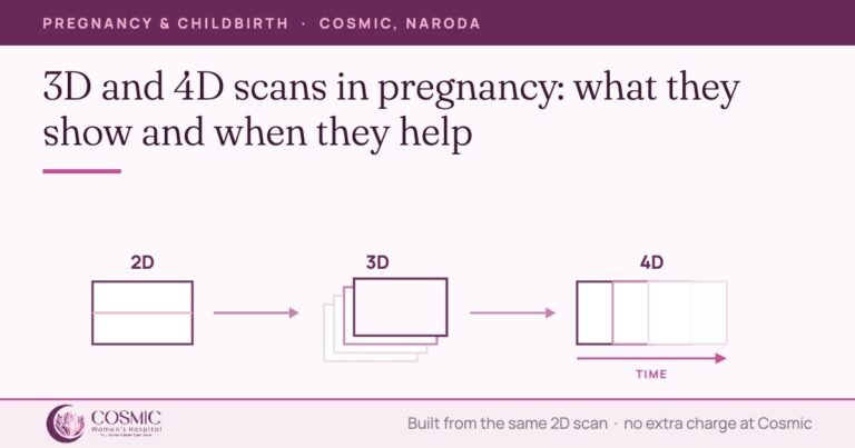
3D and 4D imaging is available at Cosmic at no additional cost. It is not sold as a separate package; it is used within the scan you are already having, when the baby’s position allows a clear view. What a 3D or 4D view adds, and where it helps most, is set out in our guide to 3D and 4D scans.
Clinically, it earns its place when there is a specific question a flat image cannot answer — most often about the face, the palate, the spine or a limb. And when it is used during a scan that is being done for a medical reason, seeing your baby’s face in three dimensions is simply part of the visit rather than an extra.
What the professional bodies advise against is a scan booked purely to produce pictures. The international society for obstetric ultrasound asks that scanning follow the ALARA principle — as low as reasonably achievable — and “not be performed solely for parental entertainment purposes”.3 The American Institute of Ultrasound in Medicine takes the same view,7 and the US Food and Drug Administration notes that keepsake images are reasonable when they come out of a scan that was medically indicated anyway.8 That is exactly how 3D and 4D are used here.
What the law requires at every pregnancy scan
The sex of the baby is never told to you. Not at any scan, not at any stage, not by any member of staff, and not for any reason. That is not hospital policy — it is criminal law under the Pre-Conception and Pre-Natal Diagnostic Techniques (Prohibition of Sex Selection) Act, 1994, and it binds the doctor, the sonologist and the family alike.
What the Act requires of a registered ultrasound facility is worth knowing, because it shapes what happens at your appointment:
- The centre must be registered before any prenatal diagnostic procedure, including ultrasound, is carried out.
- A notice stating that sex determination is not done here must be displayed prominently, in English and in the local language.
- Form F is completed for every prenatal diagnostic procedure, signed by the person conducting it, and you will be asked to give written consent.
- Records are kept for two years — the consent, the form, the images and the report.
- Disclosing the sex of the foetus “by words, signs or in any other manner whatsoever” is an offence carrying imprisonment of up to three years and a fine on a first conviction, for the doctor and for the person who asked.
So when the sonologist asks you to sign a form before the scan, that is the law working as intended, not paperwork for its own sake. Everything else on the screen — the heartbeat, the growth, the placenta, the fluid — is yours to see and ask about.
How many scans, then?
The World Health Organization recommends at least one scan before 24 weeks for every pregnant woman — to date the pregnancy, to find anomalies and multiple pregnancies, to reduce inductions for a wrongly calculated due date, and because women value it.6 That is a floor, written for the whole world including places with no ultrasound at all.
In routine practice here, a straightforward pregnancy has the 11–14 week scan and the anomaly scan, usually a dating scan before them if the dates are uncertain or there has been bleeding, and a growth scan with colour Doppler at 32 to 34 weeks, with further scans after that if anything prompts them. A high-risk pregnancy has more, and each extra scan has a stated reason attached to it. How often you are seen between the scans is set out on our maternity care page.
At Cosmic
Ultrasound and colour Doppler are done in our own department in Naroda, and both consultants perform and report the obstetric scans themselves rather than sending you elsewhere and waiting on a report. That matters most on the days it goes wrong: if a scan raises a question, the person who scanned you is the person who explains it, in the same visit.
| Item | Charge |
|---|---|
| Pelvic or obstetric ultrasound | Rs 1,200 |
| Colour Doppler | Rs 1,500 |
| 3D / 4D imaging, within a scan | No additional charge |
| New consultation — general gynaecology | Rs 500 |
| Follow-up consultation | Rs 300 |
A female attendant stays with you throughout, and you are welcome to bring your husband or a relative into the scan room. If you would prefer to be seen by a woman, that is arranged as a matter of course — see the female gynaecologist page.
Worth asking at your next scan
- What are my dates, and which scan were they set from?
- Is this scan being done for a reason, or as a routine? What is the reason?
- Was the anatomy survey complete, or does anything need to be looked at again?
- Where is the placenta lying, and does that need a repeat scan?
- If a growth scan has been advised — what are we watching for, and when is the next one?
Cosmic Women’s Hospital & IVF Center
Naroda, Ahmedabad · OPD Monday to Saturday, 10 am to 8 pm · Sunday emergency only
Emergency obstetric cover 24 hours · Telephone 77-9383-9383
Naroda, Ahmedabad · OPD Monday to Saturday, 10 am to 8 pm · Sunday emergency only
Emergency obstetric cover 24 hours · Telephone 77-9383-9383
Legal notice. Prenatal sex determination is not done at Cosmic Women’s Hospital & IVF Center and is prohibited by law under the Pre-Conception and Pre-Natal Diagnostic Techniques (Prohibition of Sex Selection) Act, 1994. Ultrasound here is performed only for medical indications. This page is general information about pregnancy scans and is not a substitute for assessment of your own pregnancy.
Sources
- International Society of Ultrasound in Obstetrics and Gynecology. ISUOG Practice Guidelines: performance of first-trimester fetal ultrasound scan. Ultrasound in Obstetrics & Gynecology 2013;41:102–113.
- ISUOG Practice Guidelines (updated): performance of 11–14-week ultrasound scan. Ultrasound in Obstetrics & Gynecology 2023;61:127–143.
- ISUOG Practice Guidelines (updated): performance of the routine mid-trimester fetal ultrasound scan. Ultrasound in Obstetrics & Gynecology 2022;59:840–856.
- UK National Screening Committee. Screening tests for you and your baby: 11 physical conditions (20-week scan). GOV.UK.
- Royal College of Obstetricians and Gynaecologists. The Investigation and Management of the Small-for-Gestational-Age Fetus. Green-top Guideline No. 31, 2nd edition, 2013 (revised 2014).
- World Health Organization. WHO recommendations on antenatal care for a positive pregnancy experience, 2016 — recommendations B.2.4 and B.2.5.
- American Institute of Ultrasound in Medicine. Prudent Use and Safety of Diagnostic Ultrasound in Pregnancy. Official Statement, 2020.
- US Food and Drug Administration. Ultrasound Imaging — fetal keepsake videos.
- Ministry of Health and Family Welfare, Government of India. Handbook on Pre-Conception & Pre-Natal Diagnostic Techniques Act, 1994 and Rules with Amendments.
Written by the clinical team at Cosmic Women's Hospital & IVF Center, Naroda, Ahmedabad.
Medically reviewed by Dr. Bhargavi Khatri — MBBS, MS Obgy (Gold Medalist), FRM. Consulting Obgyn, Infertility and IVF Specialist.
Reviewed 30 September 2026.
Medically reviewed by Dr. Bhargavi Khatri — MBBS, MS Obgy (Gold Medalist), FRM. Consulting Obgyn, Infertility and IVF Specialist.
Reviewed 30 September 2026.
Book a consultation
Tell us how we can help and we will call you back.

Cosmic Women's Hospital
Cosmic Women’s Hospital & IVF Center is a registered Level 2 ART clinic in Naroda, Ahmedabad — obstetrics, gynaecological surgery, IVF and ICSI, with ultrasound in-house and 24-hour emergency obstetric cover. Articles here are written by the clinical team and medically reviewed by Dr. Rahul Khatri or Dr. Bhargavi Khatri before publication.